 Mr. Wim van Egmond; Micropolitan Museum; Chaetoceros debilis (marine diatom), a colonial plankton organism
 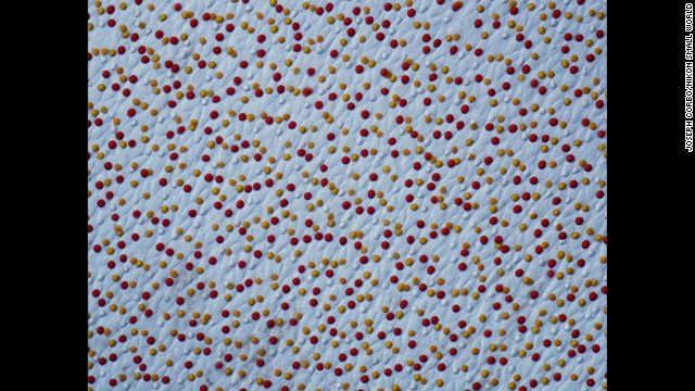 Dr. Joseph Corbo; Washington University School of Medicine; Chrysemys picta (painted turtle) retina
  Dr. Alvaro Esteves Migotto; Universidade de São Paulo, Centro de Biologia Marinha; Marine worm
  Mr. Rogelio Moreno Gill; Paramecium sp. showing the nucleus, mouth and water expulsion vacuoles
 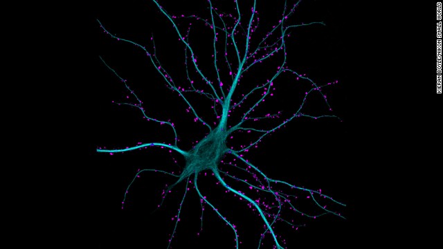 Dr. Kieran Boyle; University of Glasgow, Institute of Neuroscience and Psychology; Hippocampal neuron receiving excitatory contacts
  Miss Dorit Hockman; University of Cambridge; Chamaeleo calyptratus (veiled chameleon), embryo showing cartilage (blue) and bone (red)
  Dr. Jan Michels; Institute of Zoology, Functional Morphology and Biomechanics, Christian-Albrechts-Universität zu Kiel; Adhesive pad on a foreleg of Coccinella septempunctata (ladybird beetle)
  Ms. Magdalena Turzańska; University of Wrocław; Barbilophozia sp. (a leafy liverwort, bryophyte plant) and cyanobacteria
  Mr. Mark A. Sanders; University Imaging Centers, University of Minnesota; Insect wrapped in spider web
  Mr. Ted Kinsman; Department of Imaging and Photo Technology, Rochester Institute of Technology; Thin section of a dinosaur bone preserved in clear agate
  Miss Vitoria Tobias Santos; Universidade Federal do Rio de Janeiro, Rodrigo Evo Devo Group; Macrobrachium shrimp (ghost shrimp) eye
  Dr. Pedro Barrios-Perez; CPFC (nanofabrication), National Research Council of Canada/Information and Communication Technologies ; Silicon dioxide on polydimethylglutarimide-based resist
 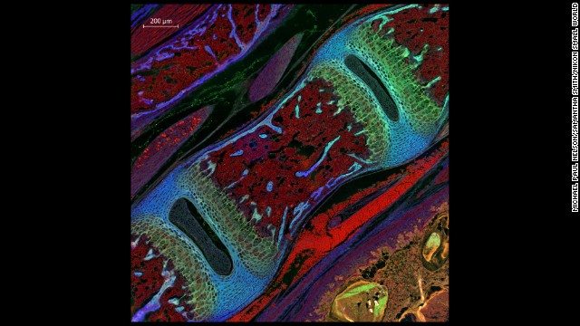 Dr. Michael Paul Nelson and Samantha Smith; Department of Pathology/Neuropathology, University of Alabama at Birmingham; Mouse vertebra section
 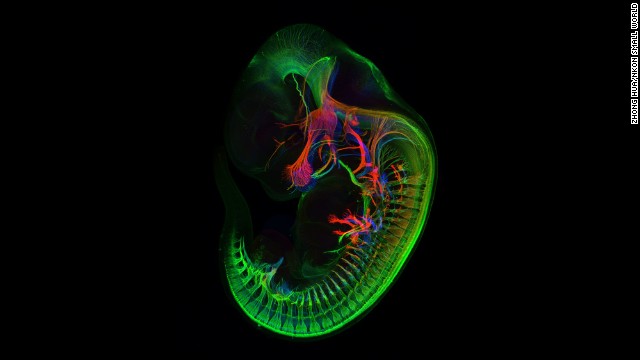 Mr. Zhong Hua; Department of Molecular Biology & Genetics, Johns Hopkins University School of Medicine; Peripheral nerves in E11.5 mouse embryo
  Dr. Christian Q. Scheckhuber; Goethe University; Podospora anserina (fungus) filamentous tip cells
 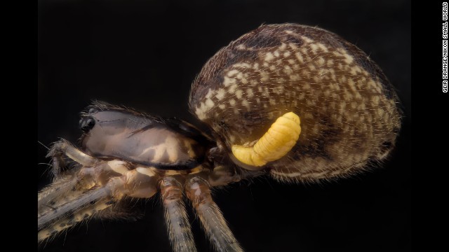 Mr. Geir Drange; Pityohyphantes phrygianus (sheet weaver spider) with a parasitic wasp larva on the abdomen
  Dr. Alexandre William Moreau; Institute of Neurology, University College London; Pyramidal neurons and their dendrites visualized in the visual cortex of a mouse brain
  Mr. Christian Sardet; Department of Life Sciences, Centre National de la Recherche Scientifique; Annelid larva
 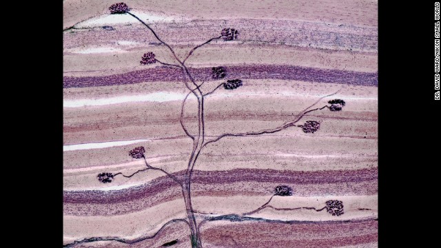 Dr. David Ward; dgward.com; Nerve and muscle thin section
  Dr. James Burchfield; The Garvan Institute; The explosive dynamics of sugar transport in fat cells
1

2

3

4

5

6

7

8

9

10

11

12

13

14

15

16

17

18

19

20
- Magnified image of marine plankton wins Nikon Small World Photomicrography Competition
- The annual contest draws entries from scientists and photographers alike
- View through the microscope is like "exploring space," says winning photographer
- Previous entries include a mosquito's heart, a flea's head and a pregnant aphid
(CNN) -- A magnified image of marine plankton has won a prestigious international photography contest for tiny works that exist, in the words of the winning photographer, in the "limbo between art and science."
Wim van Egmond, a freelance photographer from the Netherlands, took the top prize in the annual Nikon Small World Photomicrography Competition, for his magnified image of marine plankton.
It was the 11th year he had entered the contest, which draws entries from professional photographers as well as scientists, who generally produce the images in the course of their work. The winning entries are judged on their scientific and artistic merits.
PHOTOS: See high-res gallery of Nikon contest winners
"For 20 years, I've been looking through a microscope, and every time I see things I haven't seen before," said van Egmond, who has had 19 images recognized as finalists in the competition over the past decade. "It's such an endless world -- there are so many species and so many different life stages of these organisms. It's all so strange and wonderful that it's become a bit of an addiction."
The view through the microscope was like "exploring a different world, or exploring space, with these strange unknown organisms," he said.
Recognized as one of the contest's top photomicrographers, Van Egmond runs a website devoted to photomicrography called the Micropolitan Museum. He said he approached his subjects as if he were producing a portrait, trying to "capture their personalities."
"I don't invent things, I try to make it as naturalistic as possible, but these organisms are such a strange shape that it almost looks like an abstract painting," he said. "You don't have to make much of an effort to make something that is weird."
He had long been intrigued by the Chaetoceros debilis, a plant-like plankton with a corkscrew form and bristles. "It's very hard to capture because it's so 3-dimensional and so fragile. It was a bit of a challenge to make a good portrait of the organism."
The winning entry was created using software to combine various images focusing on different areas of the plankton. "The difficulty of microscopy is that when you have a ... magnification of an image, you have hardly any depth of field," he said. By combining images where some areas were in focus and others were blurred, he could create a 3-dimensional effect, he said.
Previous entries in the contest, now in its 39th year, have included a mosquito's heart, a flea's head, and a pregnant aphid.
See previous winners of the Nikon Small World Photomicrography Competition


